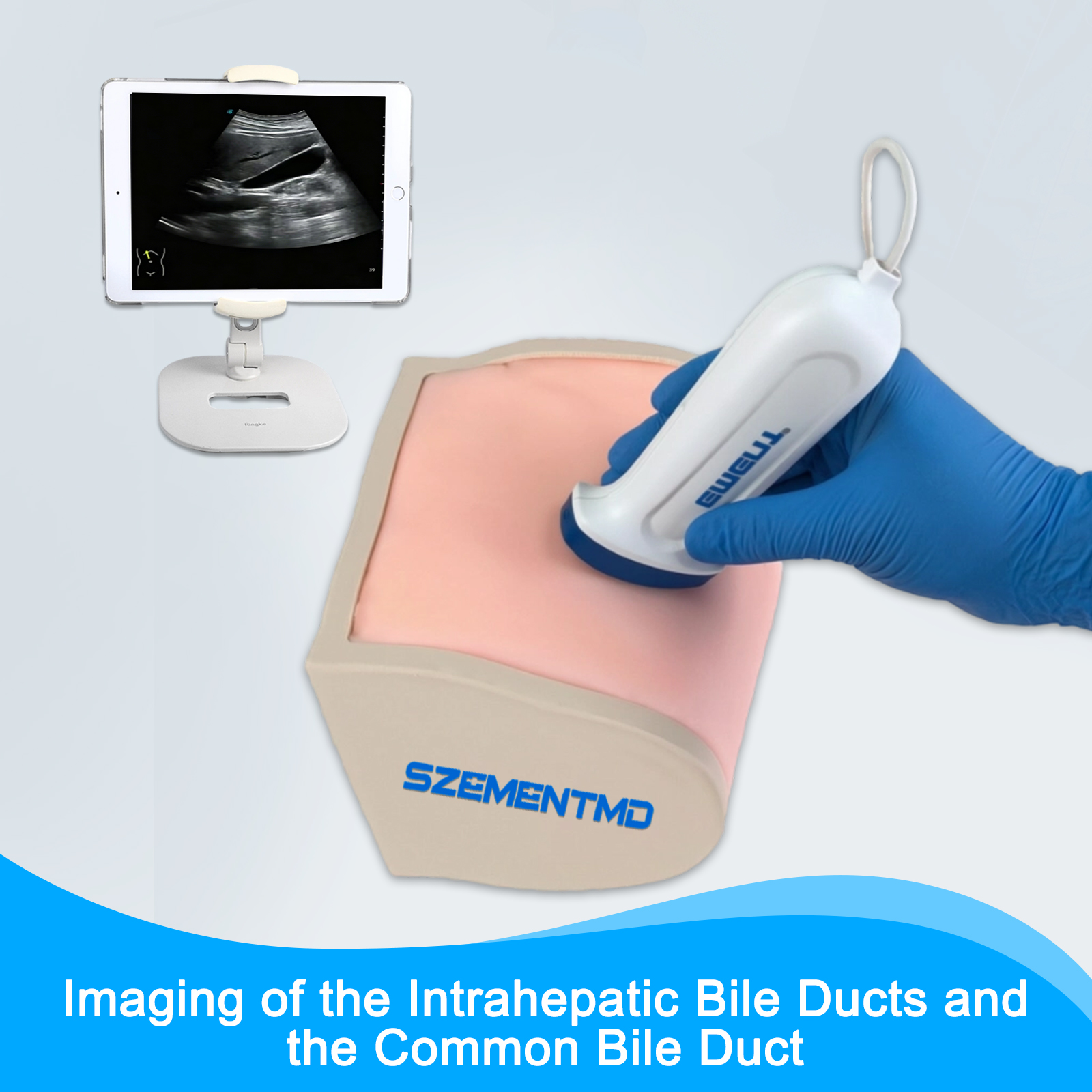
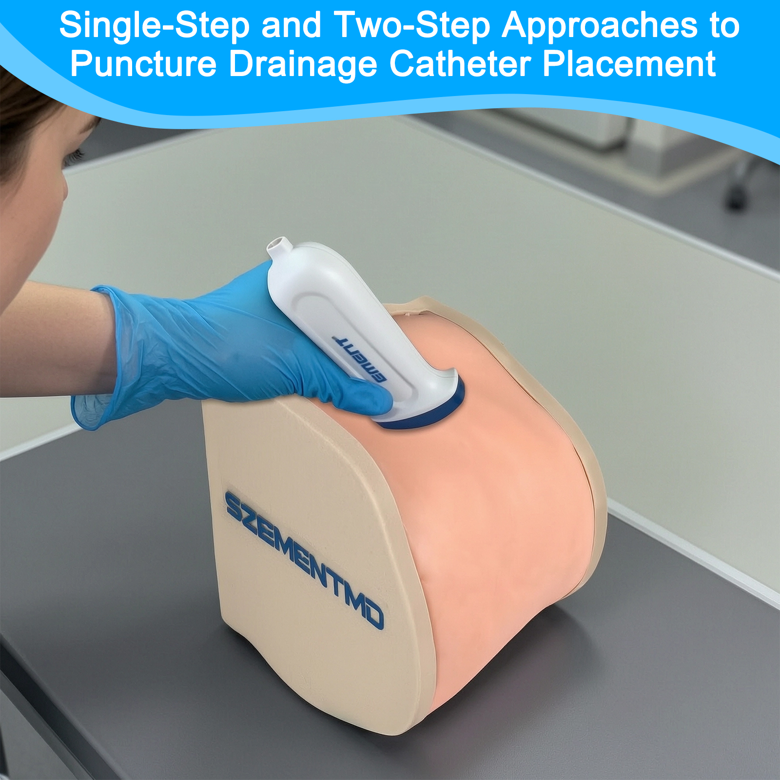
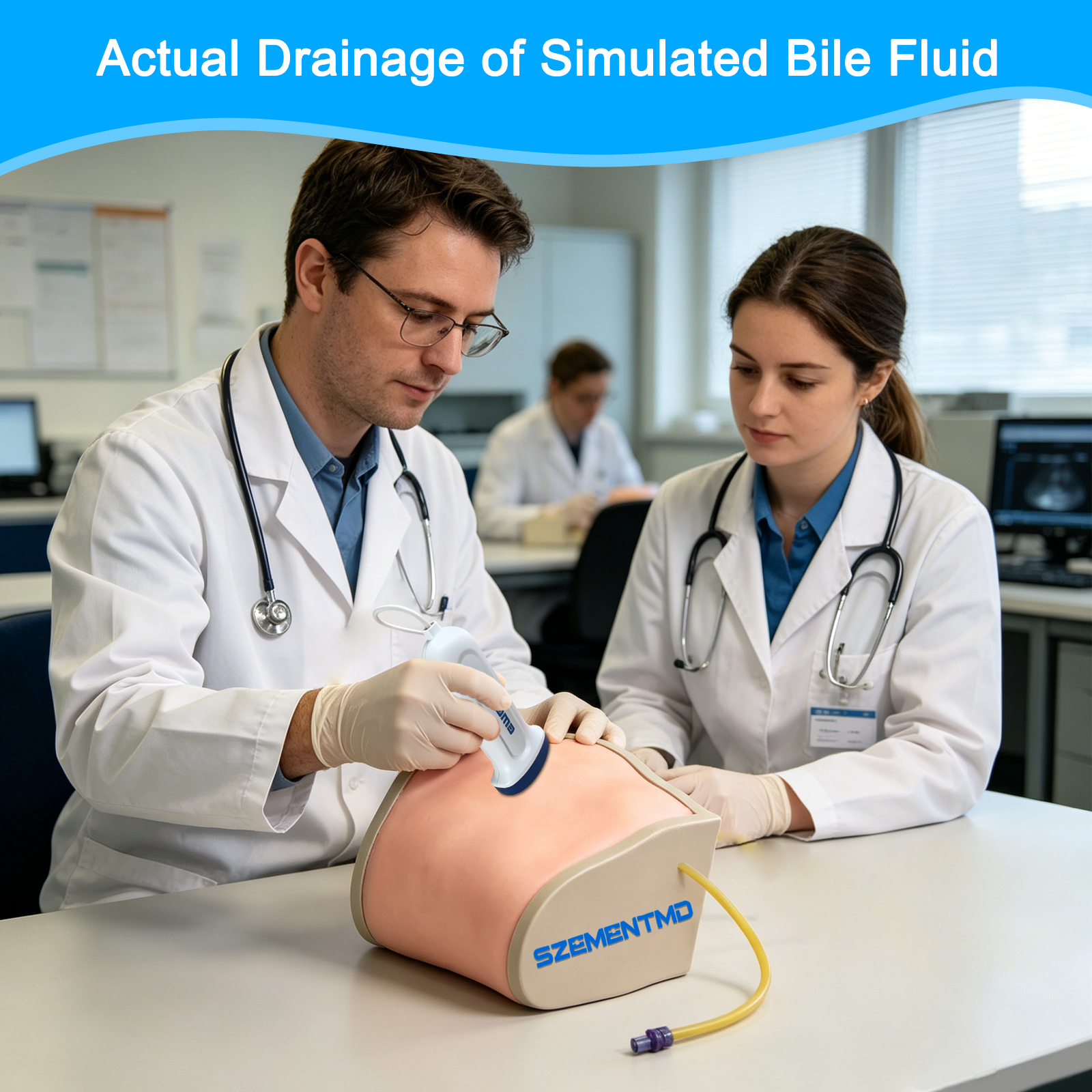
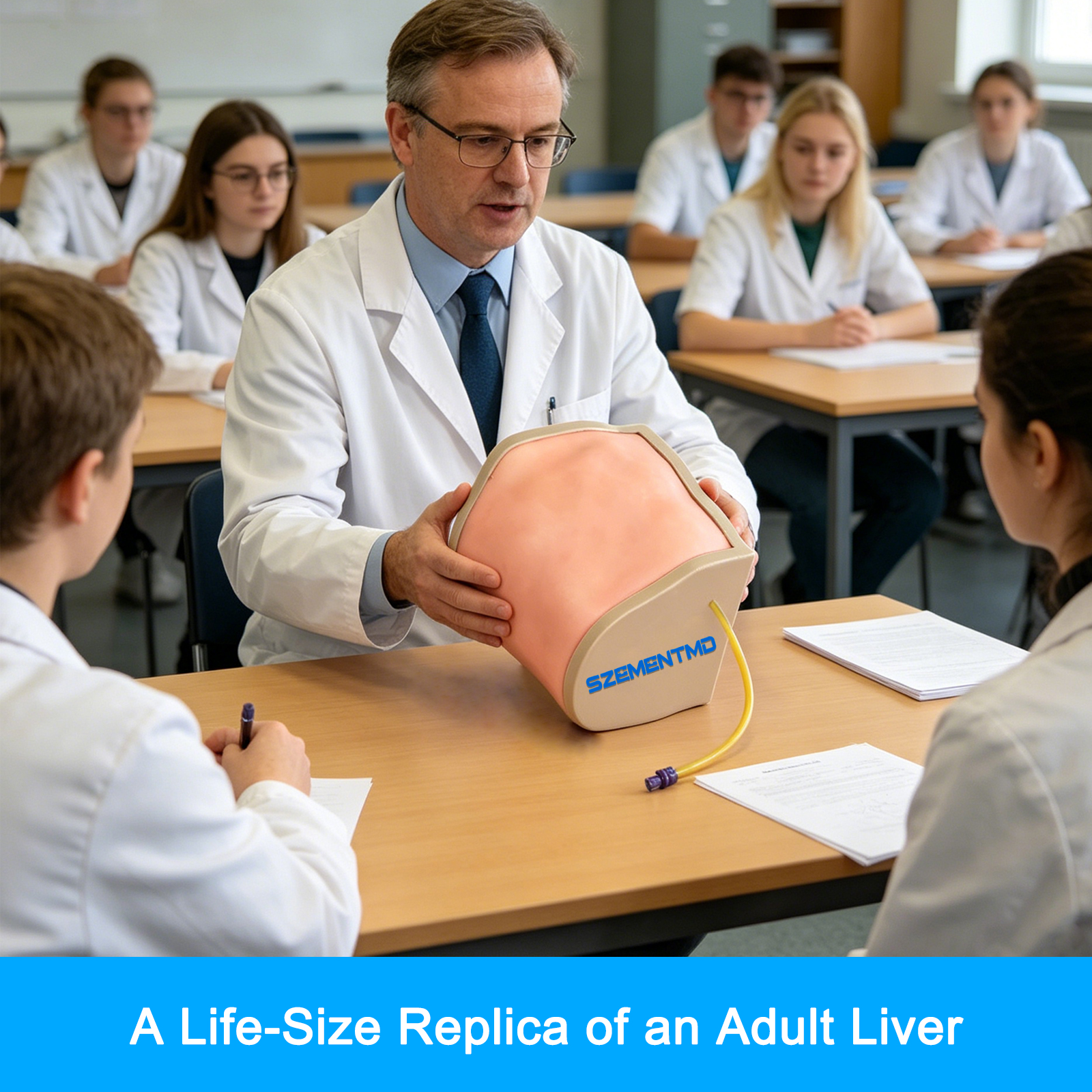
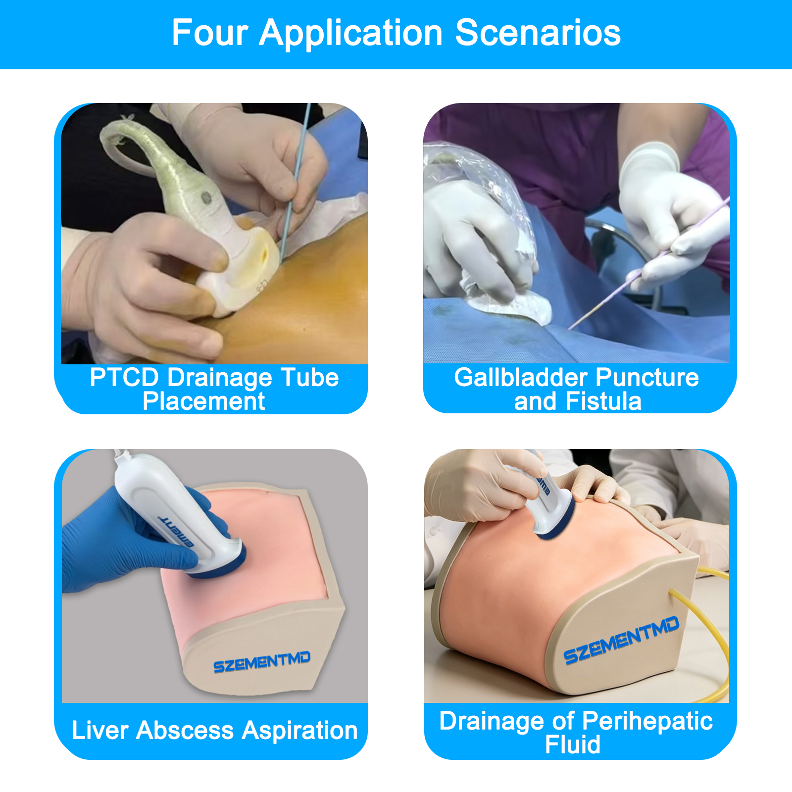
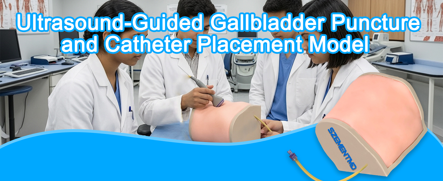
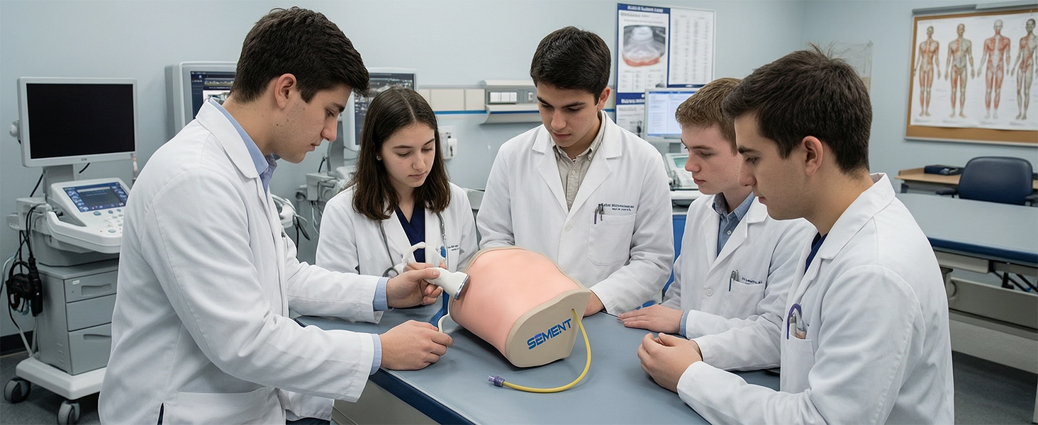
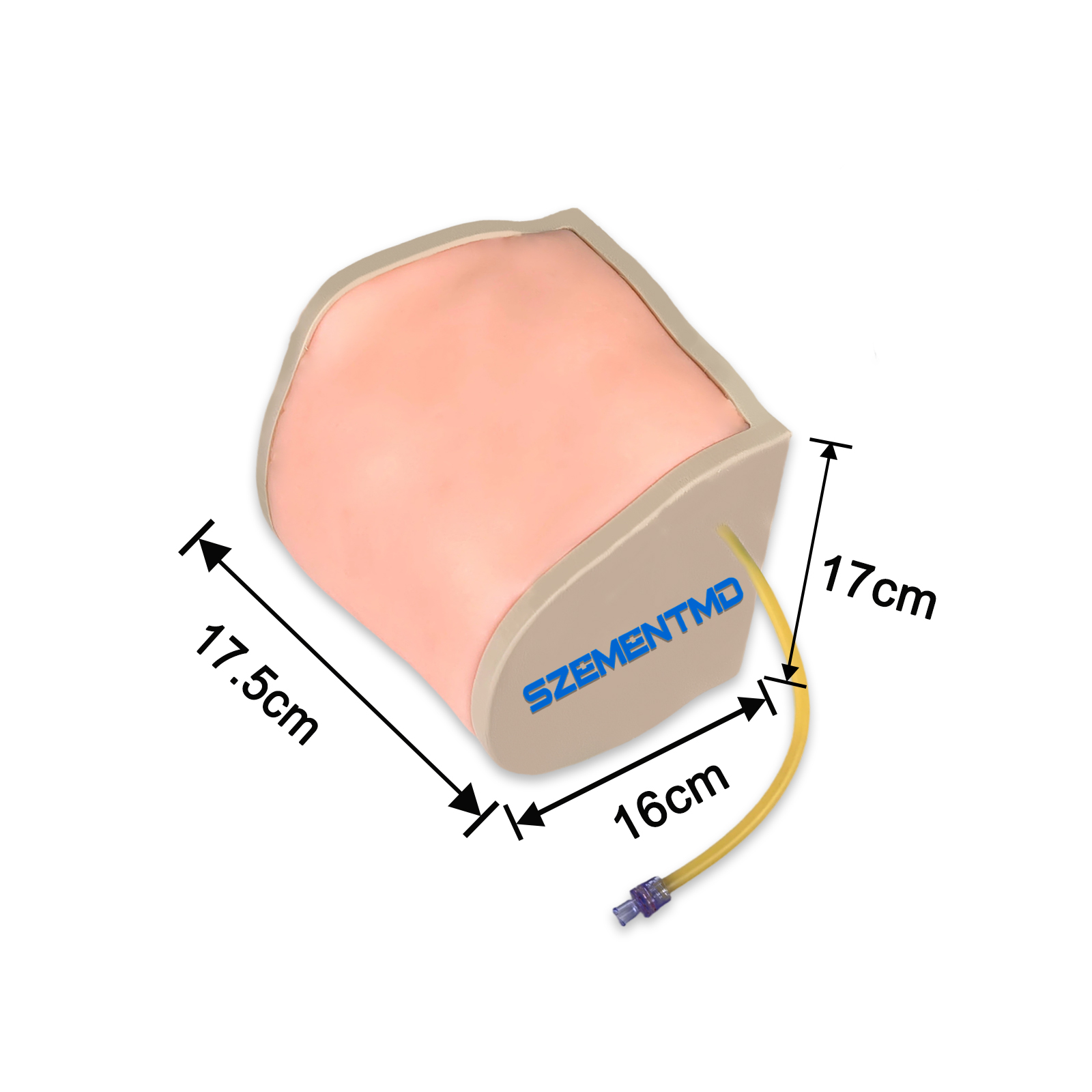
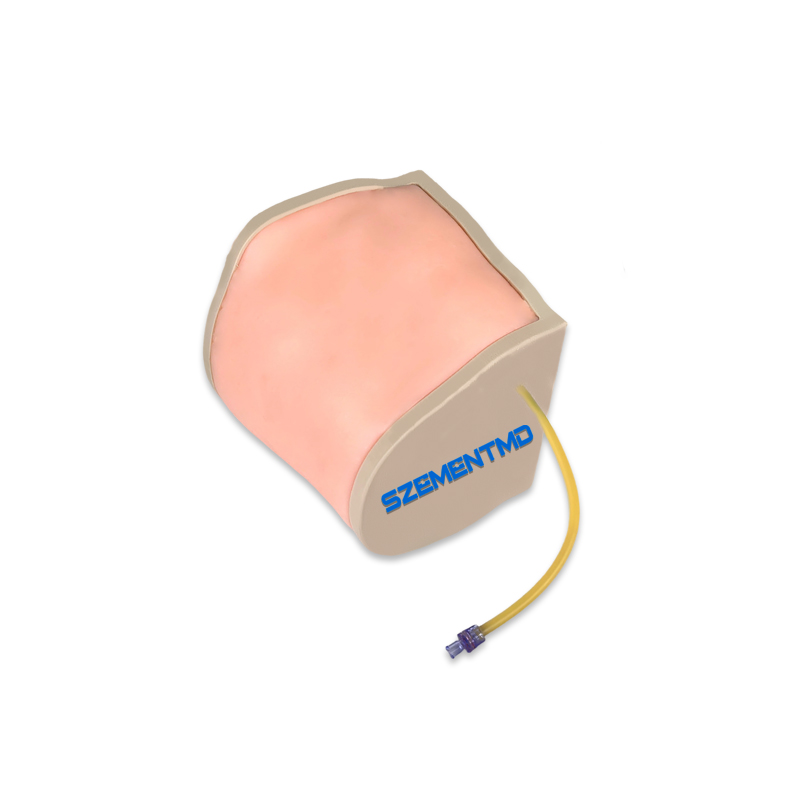
A 1:1 scale realistic simulation of an adult liver; used for imaging the intrahepatic bile ducts and common bile duct; simulates the drainage of bile fluid; Four application scenarios: 1. Percutaneous transhepatic cholangiography (PTCD) catheter placement 2. Gallbladder puncture and fistula creation 3. Liver abscess puncture 4. Periliveric fluid drainage


The key feature of the ultrasound-guided PICC placement simulator is the creation of a highly realistic, risk-free, and repeatable training platform that enables learners to safely and efficiently master this complex technique. Its key features can be summarized as follows: Highly Realistic Anatomical Structure, Laying the Foundation for Authentic Training The model is designed to closely resemble the human body both visually and tactilely. Precise Anatomical Structure: The model is designed based on real human body data. Realistic Ultrasound Images: The model is made of polymer materials with acoustic properties (density) identical to those of human tissue, ensuring that its ultrasound images are virtually indistinguishable from those of real human tissue when viewed on authentic ultrasound equipment. It clearly displays the diameter, course, and depth of blood vessels. Realistic Tactile Feedback: Puncture sensation: The resistance and “empty feel” during puncture closely resemble those of real skin tissue. Confirmation of blood return: After successfully puncturing a vessel, simulated blood return (“flashback”) is visible, providing immediate verification that the procedure was performed correctly. Dynamic simulation: High-end models can simulate arterial pulsations (via a hand-squeezed ball, etc.) and venous blood circulation, and even support Doppler ultrasound examinations. Realistic Procedural Feedback to Reinforce Skill Mastery The simulator replicates the complete clinical procedure and provides comprehensive training feedback. Supports the Complete Procedure: Enables training in the full “modified Seldinger technique,” from ultrasound guidance, vascular puncture, guidewire insertion, and skin dilation to catheter placement. Real-Time Feedback and Verification: Puncture Guidance: Enables practice of vascular puncture under ultrasound guidance. Position Verification: Verify whether the catheter tip is accurately positioned in target locations, such as the superior vena cava, using ultrasound or X-ray. Fluid Injection Verification: Verify the correct needle tip position by injecting fluid into the model. Exceptional durability and cost-effectiveness ensure long-term use Outstanding self-healing capability: Many models feature a patented self-healing material that can withstand over 1,000 repeated punctures. This material repairs itself after being punctured, leaving only a very small needle mark.
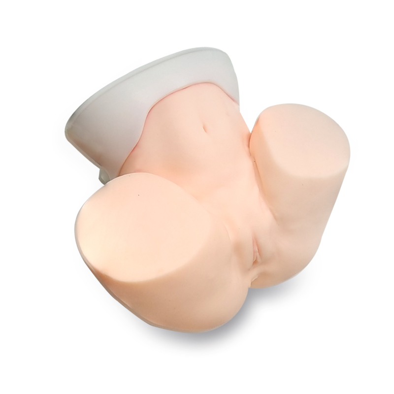
The ultrasound-guided oocyte retrieval simulator is a highly realistic training device used for assisted reproductive technology (ART) training, designed to help physicians safely and properly master the critical procedure of oocyte retrieval under transvaginal ultrasound guidance. It is described below from four core perspectives: 1. Highly Realistic Anatomical Structures and Follicle Configuration The model accurately replicates the anatomical structures of the female pelvis and reproductive system, with a particular focus on simulating the follicles—the primary target of egg retrieval procedures. Basic Anatomy: Includes complete, anatomically accurate replicas of the female abdomen, vagina, cervix, uterus, fallopian tubes, and ovaries. Some advanced models also incorporate surrounding organs such as the bladder, colon, and abdominal aorta to enhance realism and help surgeons avoid the risk of complications. Follicle Simulation: The ovarian model is designed in a sac-like shape, with multiple follicle replicas placed inside. 2. Realistic Ultrasound Imaging The model is made of specialized materials with acoustic properties highly consistent with those of human tissue, ensuring realistic images when used with actual ultrasound equipment. Imaging Quality: Clearly displays the sonographic features of the ovaries, follicles, uterus, and surrounding anatomical structures during transvaginal ultrasound. 3. Precise Oocyte Retrieval Procedure and Feedback The model is specifically designed for repeated puncture training, emphasizing realistic tactile feedback and training effectiveness. Puncture Tactile Feedback: A special material simulates the follicle wall, and a haptic feedback system replicates the resistance felt when the puncture needle penetrates the ovarian surface and follicle wall. Oocyte Retrieval Simulation: The interior of the follicles is filled with a fluid simulating follicular fluid. Some advanced models incorporate barbs and hooks to simulate real-world scenarios where oocytes adhere to the follicle wall, making aspiration difficult. During aspiration, the model also simulates the emptying and refilling of the follicles in real time. Manipulation Pathways: The needle can be moved forward and backward under ultrasound guidance, and the ultrasound probe can be rotated to obtain sagittal and transverse views. 4. Adjustable Clinical Scenarios and Complete Procedure The model supports the simulation of various clinical scenarios and covers the entire egg retrieval procedure. Variations in Ovarian Position: It can simulate complex situations, such as poor ovarian positioning caused by pelvic adhesions or anatomical variations, to train trainees to plan safe puncture paths and avoid critical structures such as the bladder, uterus, and blood vessels. Complete Procedure: The model supports the entire procedure, from ultrasound guidance (identifying the number and size of follicles) and puncture planning (avoiding surrounding organs and blood vessels) to aspiration and egg retrieval.
The ultrasound-guided uterine and adnexal model is a highly realistic simulation device used for training in gynecological ultrasound skills. It is described below from four core perspectives: 1. Highly Realistic Anatomical Structures and Pathological Findings The model accurately replicates the anatomical structure of the female pelvis and various common gynecological pathological conditions. Basic Anatomy: Includes the uterus (corpus uteri, endometrium, cervix), bilateral ovaries, fallopian tubes, bladder (including the bladder wall), broad ligament, rectum, and other landmark structures. Pathological Conditions: The model can display various pathological features, such as endometrial thickening/protrusion, uterine fibroids (simulating various sizes), ovarian cysts/follicles, large ovarian masses, pelvic effusion (in the uterorectal pouch), and abscesses. 2. Realistic Ultrasound Imaging The model is made of specialized materials (such as the patented Simulex™ technology) that closely match the acoustic properties of human tissue, ensuring realistic imaging results. Imaging Quality: Produces high-fidelity images on real ultrasound equipment, clearly displaying the ultrasonic characteristics of structures such as the uterus, ovaries, endometrium, fibroids, and cysts. Device Compatibility: Compatible with mainstream 2D, 3D, and 4D ultrasound imaging systems on the market. Training Objectives: To help trainees practice identifying normal pelvic anatomical structures and pathological sonographic patterns. 3. Repeatable Puncture and Durability The model is specifically designed for repetitive training, with an emphasis on durability and a realistic feel. Self-healing material: Key areas are made of patented, self-healing biomimetic tissue that maintains its shape and function even after multiple punctures, allowing for extensive repetitive training. Realistic feel: The resistance and tactile feedback during puncture closely mimic actual clinical procedures. 4. Multi-Pathway Operations and Clinical Scenario Adaptation The model supports multiple operational pathways and can be configured to meet different training needs. Operating Pathways: Supports both transabdominal and transvaginal ultrasound scanning pathways, enabling training in probe handling, positioning, and movement techniques for different approaches. Scenario Adaptation: Can be configured as a normal anatomical model or a model featuring specific pathological conditions. Complete Workflow: Supports a comprehensive training workflow ranging from probe operation and image optimization to anatomical structure recognition and lesion measurement.
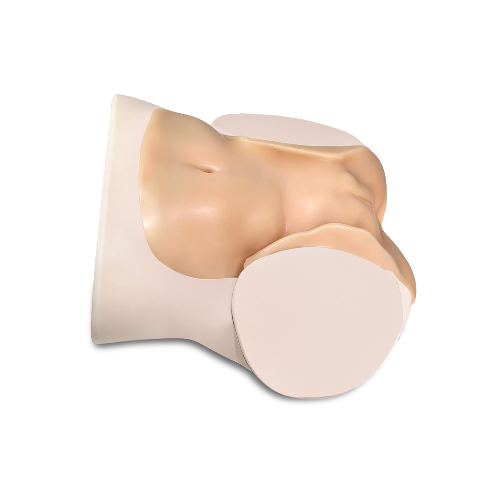
The core value of ultrasound-guided amniocentesis models lies in providing physicians with a safe, repeatable training platform to master this highly precise prenatal diagnostic technique. These models typically have the following four key features: 1. Highly Realistic Anatomical Structure Core Structure: Features a 16-week-old fetus, with approximately 200–250 ml of amniotic fluid in the uterine cavity 2. Realistic Ultrasound Imaging The model is made of a special simulation material with acoustic properties that closely mimic those of human tissue. Imaging Quality: Key structures such as the uterine wall, placenta, umbilical cord, fetus, and amniotic fluid pockets can be clearly visualized on real 2D, 3D, or 4D ultrasound equipment. Design Purpose: To help trainees practice identifying normal ultrasound anatomical structures and accurately locate target areas for puncture. 3. Repeatable Puncture Procedures The model is specifically designed for repeated puncture training, with an emphasis on durability and realistic tactile feedback. Durability and Self-Healing: Key areas are made of “self-healing” material, which maintains its shape even after multiple punctures and can withstand thousands of procedures. Realistic Tactile Feedback: The resistance felt during puncture closely mimics that of real human tissue, and the model supports staged training, such as sequentially puncturing the abdominal wall, uterus, and amniotic cavity. 4. Adjustable Clinical Scenarios To increase the difficulty and adaptability of training, the models support the simulation of various clinical conditions. Amniotic fluid volume adjustment: Using accompanying equipment, the models can simulate conditions such as polyhydramnios or oligohydramnios, training students to respond effectively to procedures of varying difficulty. Complete procedural workflow: The models support the entire procedural workflow, from ultrasound guidance and disinfection/draping to puncture and fluid aspiration; some models can even simulate complex scenarios such as twin pregnancies.
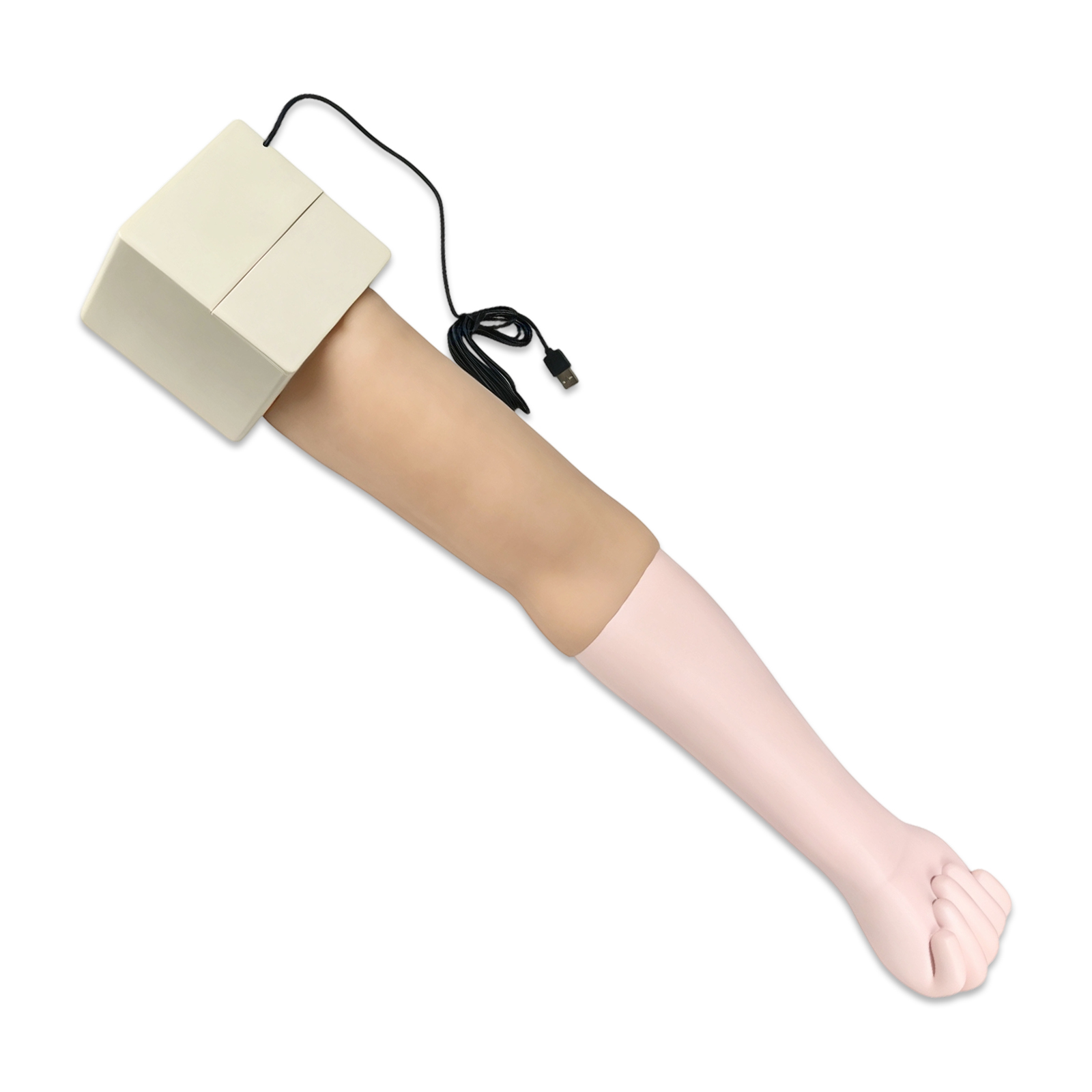
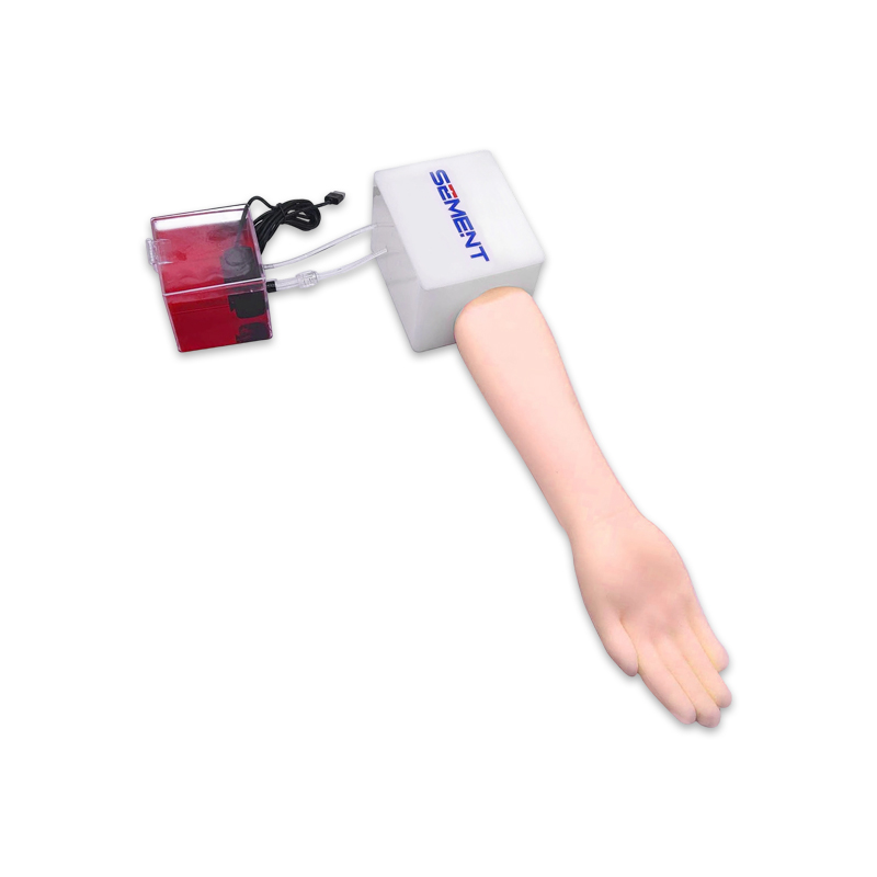
1. High-precision biomimetic anatomical structure for clear and lifelike ultrasound imaging A 1:1 reproduction of the anatomical layers of the radial artery in the forearm, crafted from medical-grade biomimetic acoustic materials. Under ultrasound scanning, vascular borders are distinct, tissue layers are clearly defined, and there is no artifact interference, highly replicating a real clinical ultrasound view. It can be used for core training in ultrasound-guided vascular identification, puncture site localization, and prediction of needle insertion angle and depth, helping operators quickly develop spatial awareness for ultrasound-guided procedures. 2. Realistic pulsation and backflow feedback, with highly realistic simulation of the entire procedure The device features a built-in simulated arterial pulsation system, supporting training for the entire procedure—including ultrasound-guided radial artery puncture and catheterization, as well as arterial blood collection. The simulated skin and blood vessels provide needle insertion resistance and a puncture sensation that closely mimic real-world clinical sensations. Upon successful puncture, clear backflow feedback is displayed, creating a highly immersive training experience that effectively shortens the clinical adaptation period for beginners. 3. Medical-grade, durable material offering excellent value for frequent use Made from high-density medical-grade simulated silicone, it is safe and odorless. It withstands repeated needle punctures and shows no visible puncture marks even after hundreds of punctures. It resists leakage and deformation, and its ultrasound visibility remains stable without degradation. The smooth surface is easy to clean and can be wiped down with medical-grade disinfectants. It is well-suited for high-frequency training scenarios in medical schools and hospitals, offering a long service life and effectively reducing the cost of training consumables. 4. User-friendly and highly adaptable design—ready for training right out of the box Featuring an ergonomic, modular design, it stands securely and is easy to operate, requiring no complicated installation or setup. It is compatible with a wide range of commonly used clinical ultrasound diagnostic devices and can be used with standard medical consumables such as conventional puncture needles, indwelling needles, and syringes. It can be flexibly placed on a desk or training table, making it suitable for various training settings, including classrooms, training labs, and clinical departments. 5. A versatile training aid for multiple scenarios and an essential tool for skill enhancement Widely applicable in various settings, including anesthesia, nursing, and emergency medicine education at medical schools; resident training and skill assessments in hospital anesthesia departments, ICUs, and emergency departments; specialized nurse training; and pre-hospital emergency response drills. This is a specialized training aid designed for standardized practice of ultrasound-guided radial artery puncture. It helps beginners quickly master the fundamentals of the procedure and is also suitable for skill reinforcement and assessment among healthcare professionals, effectively reducing risks associated with clinical procedures.

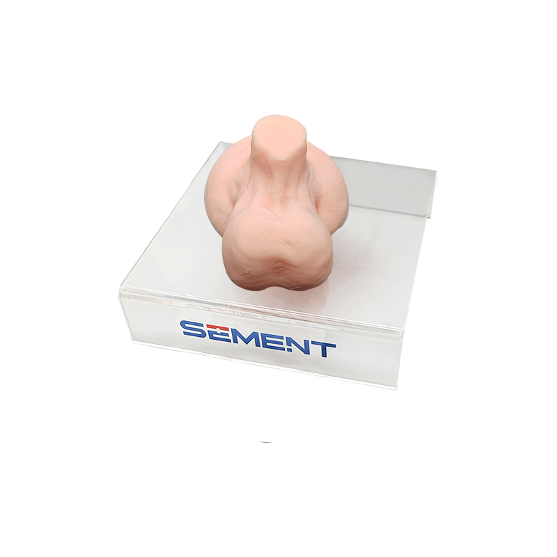
1. High-precision biomimetic anatomical structures for clear and lifelike ultrasound imaging This model faithfully reproduces key anatomical structures and tissue layers of the male testis, epididymis, and spermatic cord on a 1:1 scale. Crafted from medical-grade biomimetic materials, it produces clear and natural images under ultrasound scanning, with distinct tissue layers and no artifact interference, highly replicating a real clinical ultrasound view. It can be used for training in core skills such as anatomical identification, lesion localization, and prediction of needle insertion angle and depth under ultrasound guidance, helping operators quickly develop spatial anatomical awareness during ultrasound procedures. 2. Supports full-process interventional procedures, authentically replicating the clinical feel Supports full-process training for various interventional procedures, including ultrasound-guided testicular biopsy and drug injection; the simulation closely mimics the clinical feel of real human tissue, accurately reproducing the flexibility of the skin and soft tissues, needle insertion resistance, and the sensation of penetrating tissue. This allows users to repeatedly refine their puncture techniques and operational details, effectively shortening the clinical adaptation period for novices and improving procedural accuracy. 3. Made from durable, medical-grade material, offering excellent value for frequent use Made from high-density medical-grade silicone, this product is safe and odor-free. It withstands repeated needle punctures and remains resistant to visible puncture marks, cracking, and leakage even with long-term use. Its ultrasonic visibility remains unaffected by multiple punctures. The smooth surface is easy to clean and can be disinfected with medical-grade disinfectants. Designed for high-frequency use in hospital and academic training settings, it offers a long service life and effectively reduces the cost of training consumables. 4. A versatile training tool for multiple scenarios and an essential tool for skill enhancement Widely applicable in various settings, including urology/andrology education at medical schools, interventional ultrasound training, standardized training and skill assessments for medical professionals, preoperative simulation for clinicians, and specialized training exercises; this is a dedicated training device for standardized training in testicular ultrasound intervention skills. It helps beginners quickly master fundamental techniques while also serving to reinforce skills and conduct assessments for medical professionals, effectively reducing risks associated with clinical procedures.

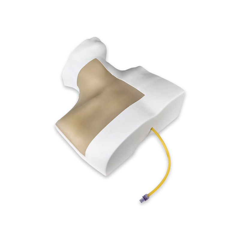
【Superior Ultrasound Imaging for Guided Training】 Engineered with ultrasound-optimized tissue that delivers exceptional acoustic transparency. The internal jugular vein, carotid artery, trachea, and clavicle are clearly distinguishable under real ultrasound devices – supports both ultrasound-guided and landmark-based blind approaches.
【Realistic Oocyte Retrieval Simulation】The Ultrasound-Guided Oocyte Aspiration Puncture Model is designed for egg retrieval and ovum collection training. It offers a high-fidelity simulation of real-life procedures, making it ideal for practicing oocyte retrieval surgery and ultrasound-guided aspiration techniques in fertility training. 【Hands-On Procedure Training】This trainer is perfect for practice of the egg retrieval process, including oocyte collection and needle insertion under ultrasound guidance. It replicates real-world surgical conditions, allowing users to refine their skills in a safe, controlled environment. 【Anatomically Accurate and Durable】With precise anatomical details, the model provides realistic practice in fertility procedures. The high-quality materials ensure durability, making the model reusable for ongoing training and simulation of retrieval and surgery techniques. 【Ideal for Fertility Specialists and Educators】Perfect for fertility professionals, medical students, and surgical trainers, this model allows hands-on experience in egg retrieval and ovum collection. It’s an essential tool for anyone looking to improve their skills in oocyte retrieval surgery. 【Comprehensive Training Tool】Use this model to practice the entire procedure from ultrasound-guided aspiration to egg retrieval. It is a reliable resource for training, ensuring students and professionals are fully prepared for real-life fertility treatments.